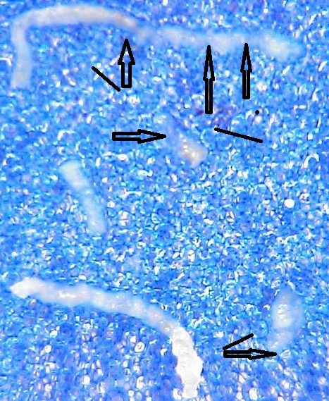
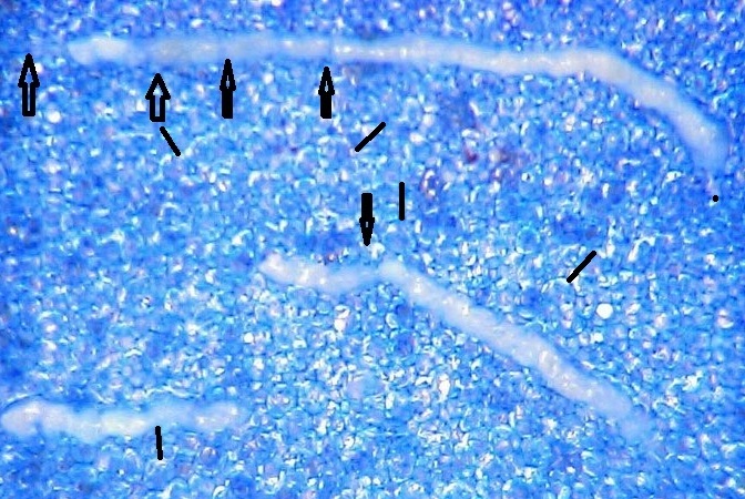
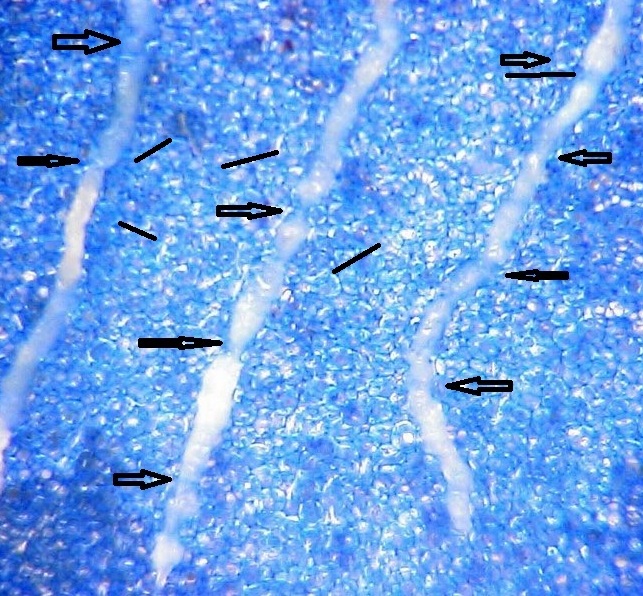
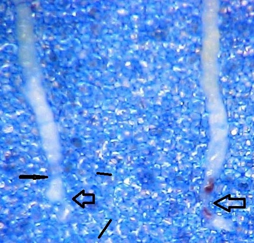
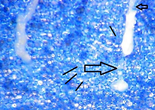
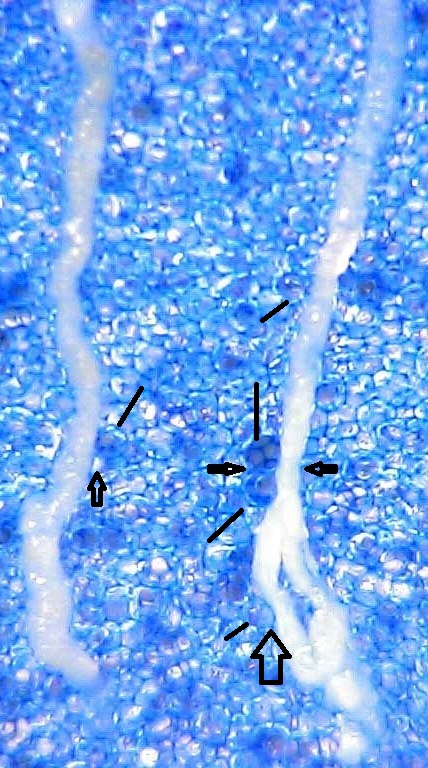
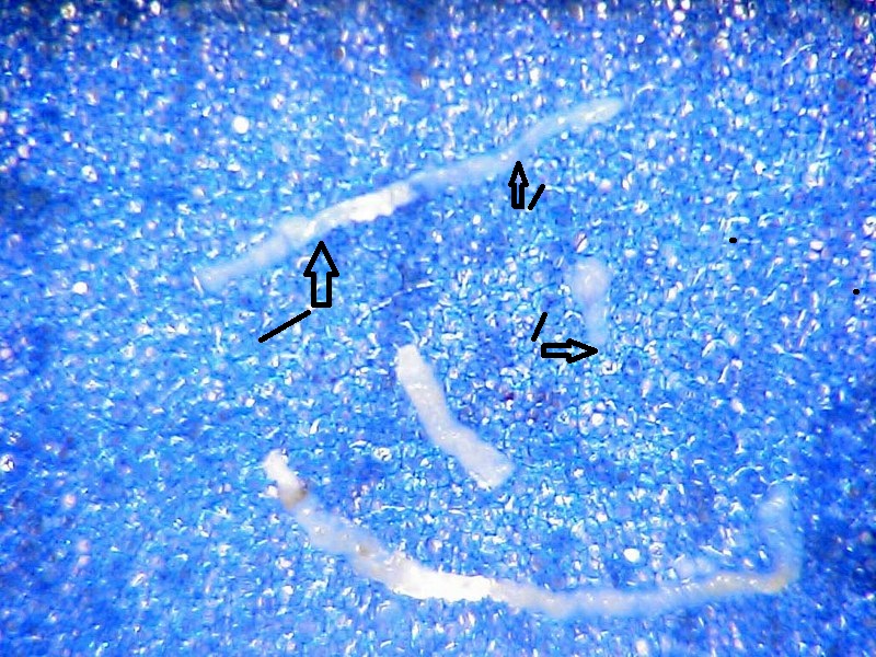
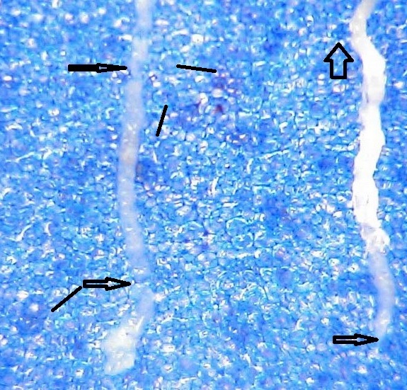
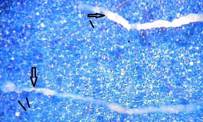
I have conducted a sort of “study” on relationship between prostate needle biopsy and the ubiquitous blue sponge at the surgical pathology laboratory. Two andomly chosen cases of prostate needle biopsy taken by an experienced urologist in the academic institution. I simply placed the cores on blue polyester pads and took regular focused pictures. The holes in the pad net are comparable with many areas of the cores, meaning that the most fragile acinar structures might be trapped in them by vacuum and pressure microwave accosted tissue processing. On the pictures, the holes are marked with lines and the core’s narrowing with arrows. Both cases show that compatibility of core’s areas and the net’s holes is not an exception.
It was impossible to publish 10 pictures in a scientific journal’s article due to space limits and perhaps the reviewers objection, but they demonstrate that the mentioned comparability of many cores’s areas with the size of the net’s holes and fragmentation happens often. The pictures look overcrowded, but informative and perhaps impressive. Two pictures were taken for a pending publication in Journal of Histotechnology.
Figures 1-4. Sextant prostate needle biopsy on 52 years old patient with elevated PSA
Figures 5-10. Figures 7-12 Sextant prostate needle biopsy on patient 62- year old with elevated PSA