Although all modern photo stand cameras on the market are able to provide a satisfactory macro image, the orientation on close-ups in gross digital pathology is a different approach in establishing connections between the gross section and the slide. This requires a special device, additionally to already available equipment, for example MacroPath D or MacroPath5 PRO, which is used during the two previously mentioned stages (mandatory specimen identification and optional general gross imaging).
This additional device is presented under the tentative name GrossSectionSnap. The proposed prototype should not be assumed as a commercially available product, although it might look as a sales pitch for a product. It is rather a direction for the development of equipment for the Implementation diagnostically oriented gross digital pathology. Any advancement cannot occur without a tool for its implementation.
Exclusive close up photography has important requirements:
intensive illumination with few shadows;
smaller depth of field to display details in focus;
sufficient immobilization to prevent specimen fluctuation due to prolonged exposure time.
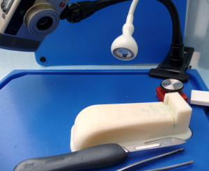
Figure 1 presents the design of a portable gross digital pathology device for close-up images. It is based on Milestone’s experimental prototype of a device for uniform and perpendicular gross sections.
Figure 1. The prototype of a portable gross digital pathology clos up image device.
A grossing board measuring 13” x 10” x 1” with a “tripod” camera holder, such as GoPro Jaws Clamp Mount, represents the basic design of the portable grossing digital pathology device for close-up images. The size and configuration of the board depends on the predominant type of specimen. Dermatopathological specimens do not need the same size of board as required by orthopedic or general surgery specimens (mastectomy, colon, lung, etc.) A waterproof LED strip is inserted into the grooves of the grossing board on it periphery in order to diminish shadows. The inserted in the board cutting platform is situated against the camera for close-up images. The camera has a ring flash for circular illumination. Depending on the type of specimen, the location of the camera and the flexible camera holder should be determined experimentally for the most favorable foreshortening in displaying optimal representative perspectives of the area the section’s shooting. The camera’s operations, including Wi-Fi variants, should be limited to a minimal number of focused foreshortened positions.
In general, the camera can work in video mode. The video option may seem attractive, but framing to choose the most appropriate image might be time consuming for a pathologist. In fact, the grossing person knows definitely the section that was placed in the cassette and the images that require the additional attention of the pathologist.
It appears that the Device for Uniform Section (DUS) is an optimal cutting platform. [1,2,3]If the size of the specimen exceeds the platform’s dimensions, which are designed for biopsies and small specimens, a different grossing platform can be developed. The modern 3D computer printer technology with availability of extrusion plastic material provides such option. As a different option, a specimen holding case, similar to a hard-pressed carton, can be used for adequate immobilization, especially in bone cutting. [4,5]
The device supposed to work in a hectic assembly line like surgical pathology laboratory environment. It should be portable and easily moved from one grossing station to another. Moreover, the device should allow easy cleanup of the mess created by grossing. GoPro type camera holders with clamps can be easily detached from the board.
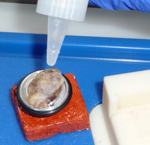
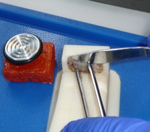
Figures 2 and 3. Frozen section diagnostic gross close-up images application
The main purpose of the device is that the pathologist can obtain a direct gross image of the most diagnostically valuable section that corresponds to the slide. The images can accompany the pathology report as part of the gross description. This design is developed from my approaches of both the pathologist and the pathologists’ assistant. I wish I had it in my work. The prototype might be an incentive for manufacturers to develop an advanced device at the level of modern optical technology.
The main purpose of the device is that the pathologist can obtain a direct gross image of the most diagnostically valuable section that corresponds to the slide. The images can accompany the pathology report as part of the gross description. This design is developed from my approaches of both as a pathologist and a pathologists’ assistant. I wish I had it in my work. The prototype might be an incentive for manufacturers to develop an advanced device at the level of modern optical technology.
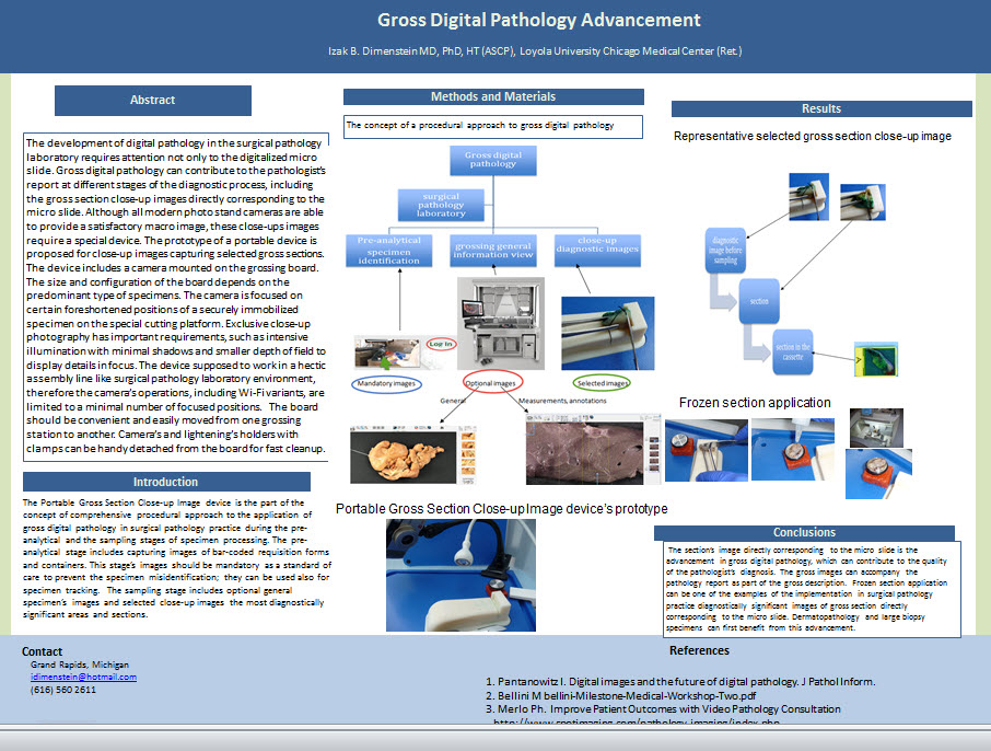
The device was presented in a poster “Gross Digital Pathology Advancement” at PathologyVision2015 in Boston in September 2015.
References
1. Dimenstein IB. Third-hand Immobilization in Gross Sectioning of Pathology Specimens. HistoLogic. 2013; June: 8-10.
2. Dimenstein IB. The concept of adjustable immobilization to aid grossing and facilitate uniform biopsy slices. Technical Note. Journal of Histotechnology. 2013; 36:106-109.
3. Dimenstein IB: Poster “The Device for Uniform and Perpendicular Sections.” Journal of Histotechnology. 2014; 37: 144.
4. Dimenstein IB: Hard Pressed Cardboard for Bone Grossing Immobilization. AAPA Newsletter. 2007;.4:25.
5. Dimenstein IB. Bone grossing techniques: helpful hints and procedures. Annals of Diagnostic Pathology. 2008; 12:191-198.


