Sampling Immobilization in Surgical Pathology

Immobilization during sampling is underestimated in grossing  (cut in) in the surgical pathology laboratory. There is no question that surgical pathology gross section requires a firm surface beneath the specimen. Though for different reasons, the firmness of the specimen determines the need for immobilization. In the vast majority of cases, it is not a problem for a skilled grossing person to make reasonably satisfactory sections. However, some specimens require certain skills supported by special techniques, devices, gadgets, and improvised at-hand materials.

Nobody is cutting in the air. It is impossible to cut on a pillow. The object of the cutting needs immobilization. Specimen sampling immobilization has three main aspects: cutting surface, holding instrument (including a hand), immobilization devise.

The importance of the cutting surface is often underestimated in grossing practice. The cork boards disappeared from practice for good. Filthy, with uneven surface, they are appropriate now for stretching a specimen in preparation for even fixation. However, they had a rational idea of additional immobilization of the specimen while cutting due to friction between the surface the outer layer of the specimen to be cut. In this regard, the modern grossing board with their smooth surface, more or less acceptable for large specimens due to their cleaning convenience, are not appropriate for biopsy and small specimen cutting because the surface should “grasp” for immobilization during cutting.

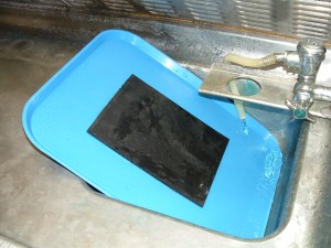
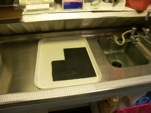
For a long time, I’m propagating the well know rubber surface of the cutting board. Rubber provides the necessary resistance to the cutting instrument, friction, and relative “softness” of the surface.  A cafeteria tray can be an improvised example of such a board.


easy to clean

easy to clean

Tray cutting board

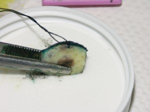
As an equivalent of the rubber surface, a Styrofoam material can be useful as a cutting surface. Because Styrofoam board re unpractical for many reason, the insert of a standard prefilled formalin container’s lid, can be an excellent “individual for every specimen cutting board.”

skin section ready

It seems that in the future manufacturer will provide the surgical pathology laboratory with gelled cut resistant surface grossing boards, like the handle of hair brush, but it will not occur tomorrow.

In the foreseeable future, nothing can substitute as a holding instrument a forceps. The industry offers a great variety of them that is justified due to different kinds of specimens and personal preferences. Among them, three types of forceps can be distinguished for practical purpose: 1. thin branch forceps; 2. Regular serrated forceps; 3. Russian holding forceps

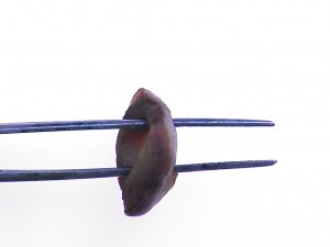
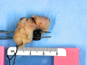
Thin branch forceps are used for different purposes in sampling biopsies. They are indispensible in sampling specimens with two open areas as in a cervical cone biopsy

LEEP

Cervical cone initial ink

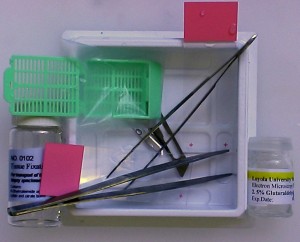
or  during triage kidney biopsies as a part of a kit.

Renal needle biopsy triadge kit

Renal needle biopsy triadge kit


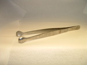
Russian holding forceps is indispensible for situation that require strong forth in specimen holding for immobilization, for instance in bone or other calcified specimen cutting of sewing.


Russian Tissue Forceps 10" fits the notch

Russian Tissue Forceps 10″ fits the notch

Russian Tissue Forceps (10" and 6")

Russian Tissue Forceps (10″ and 6″)


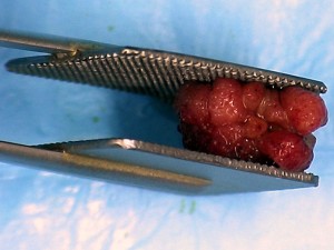
Although a paddle forceps is useful for cutting fatty tissues or sometimes thin specimens, it is especially useful for fragile large nodulated polyps. The grip of a 15 cm length with a 2.1 cm x 2.1 cm pad makes the cut easier to keep together otherwise falling apart fragments.

slide show 5


Immobilization devices are critical for successful sampling, especially in a case of calcified specimens. This is the reason that most of devices, contrivances, and gadgets are designated for bone cutting.

Our book grossing Bones:Principles, Techniques, and Instruments (2017 Amazon.com) describes bone immobilization in detail.

CoverGrosBonesAmazon


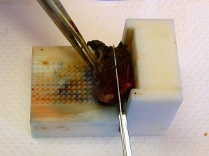
A completely different technological approach presents the Biopsy Uniform Section device which implements the principle of “third hand” immobilization. The core of the design is its three features: a specific (2 mm, 3 mm, and 4 mm) horizontal sidebar for a chosen thickness of the section, a vertical sideboard, and a V- notched slot for the cutting blade. The sideboard provides immobilization for the end of tissue to be cut functioning as the “third hand.”

BUSMilestonePrototype


Bones in the Surgical Pathology Grossing Room

Our book Grossing Bones: Principles, Techniques, and Instruments (available now on Amazon.com) summarizes specific techniques applied on different bone specimens, including tumors. The decalcification section is more comprehensive and detail oriented.

CoverGrosBonesAmazon


Table of Contents

Preface………………………………………………………………………………………………….6

General principles of bone grossing ………………………………………………………. 9

Bone gross composition ……………………………………………………………………… 10

Grossing technology terminology ………………………………………………………….14

Preservation ………………………………………………………………………………………..15

Fixation vs. in the fresh state…………………………………………………………………16

Fixation……………………………………………………………………………………………….18

Section ………………………………………………………………………………………………..21

Grossing techniques and instruments…………………………………………………….24

Orientation for gross section…………………………………………………………………25

Immobilization …………………………………………………………………………………..28

Hard-pressed cardboard packing cartons ……………………………………………..30

Other immobilization devices……………………………………………………………….35

The “third hand” principle in immobilization ………………………………………..36

Holding instruments……………………………………………………………………………39

Saws………………………………………………………………………………………………….40

Hand saws…………………………………………………………………………………………41

Power saws ………………………………………………………………………………….. ….45

Reflections on hand vs. mechanical saws …………………………………………….51

Summarization of bone grossing section manual technique…………………..53

Other instruments………………………………………………………………………….,,,55

Wrong practices ………………………………………………………………………………58

Untested challenges …………………………………………………………………………59

Decalcification …………………………………………………………………………………61

General consideration ………………………………………………………………………62

Methods of decalcification ………………………………………………………………. 63

Decalcification reagents ……………………………………………………………………64

Simultaneous fixation and decalcification ………………………………………….66

EDTA considerations ……………………………………………………………………….67

Decalcification and advanced histology studies ………………………………….68

Reflections on the choice of a decalcifier ……………………………………………69

End point of decalcification……………………………………………………………….70

Methods of end point determination………………………………………………….71

Radiography…………………………………….……………………………………………..72

Chemical method…………………………………………………………………………….72

Physical methods (bending, probing, others) …….……………………………..74

Some technical details of decalcification process……………………………….76

Agitation ………………………………………………………………………………………76

Rinsing…………………………………………………………………………………………77

Microwave decalcification ……………………………………………………………..78

Decalcification on the block, or surface decalcification …………………….79

Some recommendations from surgical pathology practice………………..80

Teeth, Nails, and Gout specimens ………………………………………………….83

Grossing techniques for selected bone specimens …………………………..85

Femoral head ………………………………………………………………………………86

Degenerative bone disease (osteoarthritis)….. ………………………………..87

Avascular necrosis………………………………………………………………………..89

Fracture………………………………………………………………………………………92

Fragile bones……………………………………………………………………………….94

Rib………………………………………………………………………………………………95

Miscellaneous fragile bones ………………………………………………………….98

Bone section from larynx …………………………………………………………….100

Maxillofacial surgery……………………………………………………………………102

Nose resection …………………………………………………………………………….102

Maxillary bone …………………………………………………………………………….102

Mandible bone……………………………………………………………………………..108

Bone tumors…………………………………………………………………………………114

General principles of grossing bone tumor specimens ……………………..114

Sampling plan of bone tumor grossing ……………………………………………115

Bone tumor specimen grossing techniques     ………………………………….117

Bone tumor specimen dissection    …………………………………………………118

The choice of a saw and immobilization    ……………………………………….119

Examples of grossing bone tumor specimens………….……………………….121

Femoral bone tumor    ………….……………………………………………………….121

Pelvic tumor    ……………………….…………………………………………………….127

Sampling bone tumor after chemotherapy …………….…….…………………129

Amputations ……………………………………….……………………………………132

Small bones …….………………………………….…………………………………… 135

Small bone fragments …………………………………………………………………..137

Bone biopsy. ………………………………………………………………………………..139

Bone marrow biopsy   …………………………………………………………………..140

Bone and cartilage…………………………………………………………………………142

Grossing for undemineralized bone processing ……………………………….143

The sample size for undemineralized bone processing………………………144

Sampling techniques ……………………………………………………………………..144

Safety issues in grossing bones ……………………………………………………… 148

Personal protection equipment …………………………………………………….. 150

The protection mask   ……………………………………………………………………151

The special place for bone specimen processing ………………………………153

Mechanical injury …………………………………………………………………………154

Grossing station for bones …………………………………………………………….155

Specialized Bone Grossing Table      ……………………………………………….157

Addendum ………………………………………………………………………………….162

Special stains and immunohistochemistry (IHC) ………………….………..162

Special stains……………………………………………………………………………… 162

Immunohistochemistry (IHC) …………………………………………………….. 164

Embedding -Microtomy sectioning rationale ……………………………….. 169

Afterword ………………………………………………………………………………… .175